|
|


 Arabic
 Bengali
 Chinese
 English
 French
 German
 Hebrew
 Hindi
Hindi Italian
 Japanese
 Korean
 Malay
Malay Polish
 Portuguese
 Spanish
 Turkish
 Ukrainian
 Vietnamese
|
HISTORY OF TECHNOLOGY, TECHNOLOGY, OBJECTS AROUND US
X-ray machine. History of invention and production
Directory / The history of technology, technology, objects around us X-ray apparatus - a set of equipment for the production and use of x-rays. It is used in medicine (radiography, fluoroscopy, radiotherapy), flaw detection. X-ray devices of a special design are used in X-ray spectral and X-ray diffraction analysis.
On November 8, 1895, Wilhelm Roentgen, professor at the University of Würzburg (Germany), wished his wife good night and went down to his laboratory to work a little more. When the wall clock struck eleven, the scientist turned off the lamp and suddenly saw a ghostly greenish glow spread on the table. It came from a glass jar containing crystals of barium platinum-cyanide. The ability of this substance to fluoresce under the action of sunlight has long been known. But usually in the dark, the glow stopped. X-ray found the source of radiation. It turned out to be a Crookes pipe that was not switched off due to inattention, which was located one and a half meters from a can of salt. The tube was under a thick cardboard cap without slots. The Crookes tube was invented about 40 years before Roentgen's observation. It was an electrovacuum tube, a source, as they said then, of "cathode rays". These rays, hitting the glass wall of the lamp, were decelerated and produced a spot of light on it, but could not escape the lamp. Noticing the radiance, Roentgen remained in the laboratory and proceeded to methodically study the unknown radiation. He installed a screen coated with barium salt at different distances from the tube. It flickered even at a distance of two meters from the tube. Unknown rays, or, as Roentgen called them Khluchi, penetrated all the obstacles that turned out to be at the scientist’s hand: a book, a board, an ebonite plate, tin foil, and even a deck of cards that had come from nowhere. All materials, previously considered opaque, became permeable to rays of unknown origin. Roentgen began to stack sheets of sheet steel: two layers, three, ten, twenty, thirty. The screen gradually began to darken and finally became completely black. A thick volume of a thousand pages did not give such an effect. From this, the professor concluded that the permeability of an object depends not so much on the thickness as on the material. When the scientist illuminated the box with a set of weights, he saw that the silhouettes of metal weights were visible much better than the faint shadow of a wooden case. Then, for comparison, he ordered to bring his double-barreled gun. Then Roentgen saw a terrible sight: the moving shadows of a living skeleton. It turned out that the bones of the hand are less transparent to the Khluchi than the surrounding soft tissues. The researcher studied the radiation he discovered for 50 days. His wife, unable to withstand the silent voluntary seclusion of her husband, burst into tears, and in order to calm her down, and at the same time demonstrate his invention to a loved one, X-ray takes an x-ray of his wife's hand. On it were visible dark silhouettes of bones, and on one of the phalanges there was a black spot of a wedding ring. Only seven weeks after the start of voluntary retreat, on December 28, 1895, Roentgen sent his 30-page manuscript "On a New Type of Rays" to the Physico-Medical Society of the University of Würzburg, adding the postscript: "Preliminary Communication."
The first work devoted to the great discovery will later turn out to be immortal: nothing in it will be either refuted or supplemented for many years. The information about Khluchi, which spread all over the world in the first week of 1896, shocked the world. The new radiation was later named "X-ray" in honor of the discoverer. Roentgen also sent his manuscript to other addresses, in particular, to his long-time colleague Professor F. Exner of the University of Vienna. He, having read the manuscript, immediately appreciated it and immediately familiarized the employees with it. Among them was the assistant E. Leher, the son of the editor of the Viennese newspaper Neue Freie Presse. He asked Exner for a text for the night, took it to his father and persuaded him to urgently put important scientific news in the room. It was given on the front page, for which they even had to stop printing presses. On the morning of January 3, 1896, Vienna heard about the sensation. The article has been reprinted by other publications. When a scientific journal came out with Roentgen's original article, the issue was snapped up in one day. Applicants for the priority of the new discovery were immediately found. Roentgen was even accused of plagiarism. Among the candidates for the championship was Professor F. Lenard, who tried to name the rays by his own name. It turned out that the first x-ray was indeed made in the USA as early as 1890. The Americans had more rights to the priority in the discovery than the same Lenard, who conducted his experiments with a Crookes tube later. But Professor Goodspeed in 1896 simply asked to be remembered that the first cathode ray photograph was taken in the laboratory of the University of Pennsylvania. After all, the true nature of these rays was established only by Roentgen. World fame, suddenly fallen on a hitherto unknown provincial scientist, led him at first into confusion. He began to avoid not only reporters, but even scientists. The professor categorically rejected the harassment of businessmen, refusing to participate in the exploitation of his discovery, from privileges, licenses, patents for his inventions, for X-ray generators improved by him. The absence of a monopoly on the production of X-ray technology has led to its rapid development throughout the world. The scientist was accused of lack of patriotism. To the offer of the Berlin Joint-Stock Electrotechnical Society, which offered a lot of money and work in well-equipped laboratories, Roentgen replied: "My invention belongs to all mankind."
After the stunning success of his discovery, Roentgen again retired to voluntary imprisonment in his laboratory. He did not pause until March 9, 1896, completed the second scientific paper on the newly discovered radiation. The third and final one - "Further Observations on the Properties of the Khluches" - was put into print on March 10, 1897. In 1904, the Englishman C. Barkla experimentally confirmed the theoretical conjecture of his compatriot J. Stokes that X-rays are of an electromagnetic nature. The X-ray region on the spectrum occupies the region between ultraviolet and gamma radiation. According to one classification, this range is from 10 ~ 5 to 10 "12 centimeters, according to another - from 10 ~ 6 to 10" 10 centimeters. The invention of the German scientist caused unexpected reactions in the world. So, in 1896, Reid, a deputy from the US state of New Jersey, proposed a bill that prohibited the use of X-rays in theatrical binoculars, so that they could not penetrate not only through clothing, but also through the flesh into the soul. And the press in Europe and America warned about the dangers of "brain photography", which allows you to read the most hidden thoughts of others. In response, some businessmen advertised their products - purses, caskets, safes, even hats - capable, according to them, of protecting their contents from terrible rays. Readers particularly responded to the information that with the help of X-rays it is possible to imprint text or a picture on the gyri of the cerebral cortex for memorization. Khluchi were credited with the ability to restore youth to the elderly and life to the dying. And also turn lead into gold. But, on the other hand, more than a thousand scientific papers and almost 1896 books on the use of X-rays in medicine were published only in the "X-ray" year of 50. Back in February 1896, V. Tonkov submitted a report to the St. Petersburg Anthropological Society on the use of X-rays for the study of the skeleton. Thus, the foundations of a new discipline, X-ray anatomy, were laid. Now it has become the foundation of modern diagnostics. A little later, A. Yanovsky began to use it for a systematic examination of patients. In a combat situation, fluoroscopy was used by the Russian doctor V. Kravchenko, who equipped an X-ray room on the Aurora cruiser. In the Battle of Tsushima, he examined the wounded sailors, finding and removing fragments from the body. Radiology helped diagnose cancer and tuberculosis in the early stages. X-ray radiation in large doses is harmful to the human body. But, nevertheless, it is used to combat malignant tumors. At the beginning of the XX century. X-rays required exposure for 1,5-2 hours due to the imperfection of the equipment and the low sensitivity of the film. Then they began to use intensifying screens for shooting, between which the film was located. This made it possible to reduce the exposure time tenfold without increasing the film sensitivity. Thanks to this, radiography surpassed fluoroscopy in terms of resolution. Since X-ray film required a large amount of silver, X-ray photography gradually began to be replaced by fluorography - photography from a fluorescent screen. The fluorogram has only one light-sensitive layer and is 10-20 times smaller in area than a standard radiograph, which gives a great saving of silver while reducing radiation exposure. The image is enlarged with the help of projectors. A compact fluorographic camera mounted on an electro-optical amplifier of a stationary device makes it possible to obtain multiple images with a short interval according to a given program. This way you can register fast processes. In particular, this method is used to control the movement of a special mass containing barium (clearly visible in x-rays) through the human gastrointestinal tract. To save the film, a special selenium plate is used that accumulates an electrostatic charge. Under the influence of X-rays, it loses its charge, retaining it only in dark areas. As a result, a latent image appears on the surface of the plate. It is developed by dusting it with a finely dispersed coloring powder that accurately reproduces the distribution of light and shadows. One selenium plate withstands 2-3 thousand procedures, saving up to 3 kg of silver. The image is not inferior in quality to the radiograph.
In addition to black and white, there is color radiography. First, a color X-ray was obtained by shooting the object three times with rays of unequal hardness. In this way, three negatives were obtained, which were stained in blue, green and red, after which they were combined and imprinted on color film. Later, to reduce the radiation dose, the method of tone separation was used. A one-time exposure was needed here. Different density zones were identified in the image, and a copy of the X-ray pattern was made for each. Then they were combined on a color film, obtaining a conventionally colored image. Conventional x-rays only produce a flat image. Often this does not allow determining, for example, the exact location of a foreign body in the body, and several radiographs taken from different positions give only an approximate idea of this. Stereoradiography is used to transform a flat image into a three-dimensional one. For this purpose, two photographs are taken that make up a stereo pair: they depict the same picture, but imprinted as it is seen by the right and left eyes. When considering both negatives in a special apparatus, they are combined into one, forming a depth. With stereofluoroscopy, the patient is translucent with two tubes, which turn on alternately at a speed of 50 times per second each. Both series of pulses are fed to an image converter, from where they are alternately, synchronously with the operation of the tubes, removed by two television systems. Both pictures are combined into one with the help of polarized glasses. The depth, spatial structure, shape and size of pathological formations are also evaluated by simpler means, for example, using tomography - layered images. During the tomography, the patient lies on the table. An X-ray cutting moves above it, and a film moves under it in the opposite direction. Only those elements that are on the axis of rotation of the lever connecting the tube and the film turn out to be sharp. A series of images are taken showing thin layers a few millimeters thick. It is easy to establish from them where the foreign body or painful focus is located. With the advent of electronic computers and computers, it became possible to programmatically control the entire procedure of X-ray diagnostics - from taking pictures to taking pictures. The range of applications of X-rays is wide. In the 20-30s of the last century, radiation genetics and breeding appeared, which made it possible to obtain resistant variants of microbes with the desired properties, plant varieties with increased productivity. By exposing organisms to penetrating radiation and then by selecting them, scientists carry out accelerated biological evolution. In 1912, in Munich, M. von Laue put forward the idea of investigating the internal structure of a crystal with the help of Khluchi. His idea caused controversy among colleagues, and in order to resolve them, V. Friedrich placed a crystal in the path of the rays and next to it, on the side, a photographic plate for recording them when they deviate at a right angle, as in ordinary diffraction. There were no results until P. Knipping put the plate not on the side, but behind the crystal. A symmetrical pattern of dark spots appeared on it. This is how X-ray diffraction analysis was born. At first, its use was limited to obtaining Lauegrams - images that reflected the structure of a single crystal. They made it possible to detect lattice defects, internal stresses, etc. In 1916, P. Debye and P. Scherrer adapted this method to study polycrystalline materials - powders, alloys. Such pictures are called debyegrams. They determine the structure and composition of the samples, the size and orientation of the inclusions. In the 1930s, English scientists D. Bernal and D. Crowfoot-Hodgkin carried out X-ray diffraction analysis of proteins. The shooting revealed their internal orderliness. Thanks to this analysis, the spatial model of DNA, which was proposed in 1953 by D. Watson and F. Crick, became possible. To do this, they used the diffraction patterns of DNA obtained by M. Wilkins. X-rays are used to control the quality of various materials and products. They allow you to see internal defects - cracks, shells, lack of penetration, inclusions. This method is called X-ray flaw detection. X-rays allow art historians to look under the top layer of paintings, sometimes helping to reveal centuries-old images. So, when studying Rembrandt's painting "Danae", the original version of the canvas was discovered, later redone by the author. Many paintings in various art galleries have undergone similar research.
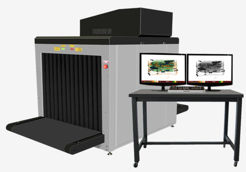
X-ray radiation is used in introscopes - devices that are now equipped with customs, checkpoints. They allow you to detect hidden explosives, weapons and drugs. Author: Pristinsky V.L.
▪ Teflon ▪ Frisbee
Artificial leather for touch emulation
15.04.2024 Petgugu Global cat litter
15.04.2024 The attractiveness of caring men
14.04.2024
▪ Drawing color pictures with white light ▪ Gel that allows you to stick sensors to internal organs ▪ Smart clothes that track posture and movement ▪ Movement of self-driving cars without marking
▪ section of the site RF power amplifiers. Article selection ▪ article Better death, but with glory, than inglorious days of shame. Popular expression ▪ article What is a sandwich made of? Detailed answer ▪ article Bicycle on skis. Personal transport ▪ article Tar varnish. Simple recipes and tips
Home page | Library | Articles | Website map | Site Reviews
www.diagram.com.ua |


 See other articles Section