|
|


 Arabic
 Bengali
 Chinese
 English
 French
 German
 Hebrew
 Hindi
Hindi Italian
 Japanese
 Korean
 Malay
Malay Polish
 Portuguese
 Spanish
 Turkish
 Ukrainian
 Vietnamese
|
VISUAL (OPTICAL) ILLUSIONS
Brief information about the structure of the eye and visual sensations. Encyclopedia of visual illusions
At leisure / Visual (optical) illusions << Back: Table of contents >> Forward: Disadvantages and defects of vision The human eye is an almost spherical body that rests in a bony cranial cavity, open on one side. On fig. 1 shows a section of the eyeball and shows the main details of the eye.
The main part of the eyeball from the outside is limited by a three-layer shell. The outer hard shell is called the sclera (Greek for hardness) or protein shell. It covers the inner contents of the eye from all sides and is opaque throughout its entire length except for the front. Here the sclera protrudes forward, is completely transparent and is called the cornea. Adjacent to the sclera is the choroid, which is full of blood vessels. In the anterior part of the eye, where the sclera passes into the cornea, the choroid thickens, departs at an angle from the sclera and goes to the middle of the anterior chamber, forming the transverse iris. If the back of the iris is colored only black, the eyes appear blue, the blackness shines through the skin with a bluish tint, like the veins on the hands. If there are other colored inclusions, which also depends on the amount of black colored substance, then the eye seems to us greenish, gray and brown, etc. When there is no colored substance in the iris (as, for example, in white rabbits), then it it seems to us red from the blood contained in the blood vessels penetrating it. In this case, the eyes are poorly protected from light - they suffer from photophobia (albinism), but in the dark they are superior in visual acuity to eyes with a dark color. The iris separates the anterior convex segment of the eye from the rest of the eye and has an opening called the pupil. The pupil of the eye itself is black for the same reason as the windows of a neighboring house in daylight, which appear black to us, because the light that has passed through them from the outside hardly comes back. The pupil passes a certain amount of light into the eye in each case. The pupil increases and decreases independently of our will, but depending on the lighting conditions. The phenomenon of adaptation of the eye to the brightness of the visual field is called adaptation. However, the main role in the process of adaptation is played not by the pupil, but by the retina. The retina is the third, inner shell, which is a light and color-sensitive layer. Despite its small thickness, it has a very complex and multilayered structure. The light-sensitive part of the retina consists of nerve elements enclosed in a special tissue that supports them. The light sensitivity of the retina is not the same throughout its entire length. In the part opposite the pupil and slightly above the optic nerve, it has the greatest sensitivity, but closer to the pupil it becomes less and less sensitive and, finally, immediately turns into a thin shell covering the inside of the iris. The retina is a branching of nerve fibers along the bottom of the eye, which then intertwine with each other and form the optic nerve, which communicates with the human brain. There are two types of nerve fiber endings lining the retina: some are stalk-like and relatively long, called rods, others, shorter and thicker, are called cones. There are about 130 million rods and 7 million cones on the retina. Both rods and cones are very small and are only visible at 150-200 times magnification under a microscope: the thickness of the rods is about 2 microns (0,002 mm), and the cones are 6-7 microns. In the most light-sensitive part of the retina opposite the pupil, there are almost only cones, their density here reaches 100000 per 1 mm2, and every two or three light-sensitive elements are connected directly to nerve fibers. Here is the so-called central fossa with a diameter of 0,4 mm. As a result, the eye has the ability to distinguish the smallest details only in the center of the field of view, limited by an angle of 1 °.3. So, for example, experienced grinders distinguish gaps of 0,6 microns, while usually a person is able to notice a gap of 10 microns. The area closest to the central fossa, the so-called yellow spot, has an angular extent of 6-8 °. The rods are located within the entire retina, and their highest concentration is observed in the zone displaced by 10-12 ° from the center. Here, one fiber of the optic nerve accounts for several tens and even hundreds of rods. The peripheral part of the retina serves for general visual orientation in space. With the help of a special eye mirror proposed by G. Helmholtz, one can see a second white spot on the retina. This spot is located on the site of the optic nerve trunk, and since there are no longer any cones or rods, this area of the retina is not sensitive to light and is therefore called the blind spot. The blind spot of the retina has a diameter of 1,88 mm, which corresponds to 6° in terms of visual angle. This means that a person from a distance of 1 m may not see an object with a diameter of about 10 cm if the image of this object is projected onto a blind spot. Rods and cones differ in their functions: rods are highly sensitive, but do not "distinguish" colors and are devices for twilight vision, that is, vision in low light; cones are sensitive to colors, but are less sensitive to light and therefore are daytime vision devices. In many animals, behind the retina is a thin shimmering mirror layer that enhances the effect of light entering the eye by reflection. The eyes of such animals shine in the dark like hot coals. This is not about complete darkness, where this phenomenon, of course, will not be observed. Vision adaptation is the complex process of switching the eye from cone to rod (dark adaptation) or vice versa (light adaptation). At the same time, the processes of changing the concentration of light-sensitive elements in the retinal cells, when its sensitivity increases by tens of thousands of times during dark adaptation, as well as other changes in the properties of the retina in various phases of adaptation, remain unknown. The actual data of the adaptation process are defined quite strictly and can be given here. So, in the process of dark adaptation, the sensitivity of the eye to light first increases rapidly, and this lasts about 25-40 minutes, and the time depends on the level of initial adaptation. With a long stay in the dark, the sensitivity of the eye to light increases 50000 times and reaches the absolute light threshold. Expressing the absolute threshold in lux of illumination on the pupil, an average value of the order of 10-9 lux is obtained. This means, roughly speaking, that in conditions of complete darkness, the observer could notice the light from one stearin candle, removed from him at a distance of 30 km. The higher the brightness of the initial adaptation field, the slower the eye adapts to darkness, and in these cases the concept of relative sensitivity thresholds is used. During the reverse transition from darkness to light, the process of adaptation to the restoration of some "constant" sensitivity lasts only 5-8 minutes, and the sensitivity changes only 20-40 times. Thus, adaptation is not just a change in the diameter of the pupil, but also complex processes on the retina and in areas of the cerebral cortex connected with it through the optic nerve. Immediately behind the pupil of the eye is a completely transparent, elastic body, enclosed in a special bag attached to the iris by a system of muscle fibers. This body has the form of a collective biconvex lens and is called the lens. The purpose of the lens is to refract light rays and give a clear and distinct image of objects in the field of view on the retina of the eye. It should be noted that, in addition to the lens, both the cornea and the internal cavities of the eye, filled with media with refractive indices different from unity, take part in the formation of an image on the retina. The refractive power of the entire eye as a whole, as well as individual parts of its optical system, depends on the radii of the surfaces limiting them, on the refractive indices of substances and the mutual distance between them. All these values for different eyes have different values, therefore, the optical data of different eyes are different. In this regard, the concept of a schematic or reduced (reduced) eye is introduced, in which: the radius of curvature of the refractive surface is 5,73 mm, the refractive index is 1,336, the length of the eye is 22,78 mm, the front focal length is 17,054 mm, the rear focal length is 22,78 mm . The lens of the eye forms on the retina (just like a camera lens on a matte plate) an inverted image of the objects we look at. This is easy to verify. Take a piece of thick paper or a postcard and poke a small hole in it with a pin. Then we put the pin head up at a distance of 2-3 cm from the eye and look with this eye through a hole in the paper, set at a distance of 4-5 cm, at the bright daytime sky or at a lamp in a milk flask. If the distances between the eye and the pin, the pin and the paper, which are favorable for the given eye, are selected, then in the light hole we will see what is shown in Fig. 2. The shadow of the pin on the retina will be straight, but the image of the pin will appear upside down to us. Any movement of the pin to the side will be perceived by us as a movement of its image in the opposite direction. The outline of the pinhead, which is not very clear, will appear to be on the other side of the sheet of paper.
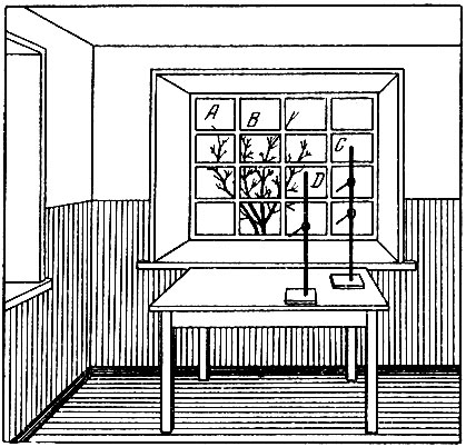
The same experiment can be done in a different way. If three holes are pierced in a piece of thick paper, located at the vertices of an equilateral triangle with sides approximately equal to 1,5-2 mm, and then the pin and paper are placed in front of the eye, as before, then three reverse images of the pin will be visible. These three images are formed due to the fact that the light rays passing through each of the holes do not intersect, since the holes are in the anterior focal plane of the lens. Each beam gives a direct shadow on the retina, and each shadow is perceived by us as an inverted image. If we put paper with three holes to the eye, and paper with one hole to the light source, then our eye will see an inverted triangle. All this convincingly proves that our eye perceives all objects in a direct form, because the mind inverts their images obtained on the retina. Back in the early 20s, the American A. Stratton and in 1961 the professor at the California Institute, Dr. Irwin Mood, set up an interesting experiment on themselves. In particular, I. Mud put on special glasses that fit tightly to his face, through which he saw everything as on the frosted glass of a camera. For eight days, walking several dozen steps, he felt symptoms of seasickness, confused the left side with the right, top and bottom. And then, although the glasses were still in front of my eyes, I again saw everything as all people see. The scientist regained freedom of movement and the ability to quickly orientate himself. In his glasses, he rode a motorcycle through the busiest streets of Los Angeles, drove a car, piloted a plane. And then Mood took off his glasses - and the world around him turned upside down again. I had to wait a few more days until everything went back to normal. The experiment once again confirmed that the images perceived by vision do not enter the brain in the same way as they are transmitted to the retina by the optical system of the eye. Vision is a complex psychological process, visual impressions are consistent with the signals received by other senses. It takes time before this whole complex system is set up and begins to function normally. It is this process that occurs with newborns, who at first see everything upside down and only after some time begin to perceive visual sensations correctly. Since the retina is not a flat screen, but rather spherical, the image on it will not be flat. However, we do not notice this in the process of visual perception, since our mind helps us to perceive objects as they really are. The bag in which the lens is fixed is a ring-shaped muscle. This muscle can be in a state of tension, which causes the lens to take the least curved shape. When the tension of this muscle decreases, the lens, under the action of elastic forces, increases its curvature. When the lens is stretched, it gives a sharp image of objects located at great distances on the retina of the eye; when it is not stretched and the curvature of its surfaces is great, then a sharp image of nearby objects is obtained on the retina of the eye. The change in the curvature of the lens and the adaptation of the eye to a clear perception of far and near objects is another very important property of the eye, which is called accommodation. The phenomenon of accommodation is easy to observe as follows: we look with one eye along a stretched long thread. At the same time, wanting to see near and far sections of the thread, we will change the curvature of the lens surfaces. Note that at a distance of up to 4 cm from the eye, the thread is not visible at all; only starting from 10-15 cm do we see it clearly and well. This distance is different for people young and old, for nearsighted and farsighted, and for the first it is less, and for the second it is more. Finally, the part of the thread farthest from us, clearly visible under given conditions, will also be differently removed for these people. Nearsighted people will not see the thread beyond 3 m. It turns out, for example, that for viewing the same printed text, different people will have different distances of best vision. The distance of best vision, at which the normal eye experiences the least stress when looking at the details of an object, is 25-30 cm. The space between the cornea and the lens is known as the anterior chamber of the eye. This chamber is filled with a gelatinous transparent liquid. The entire interior of the eye between the lens and the optic nerve is filled with a somewhat different kind of vitreous body. Being a transparent and refractive medium, this vitreous body at the same time helps to maintain the shape of the eyeball. In conclusion to his book "On Flying Saucers", the American astronomer D. Menzel writes: "In any case, remember that flying saucers: 1) really exist; 2) they were seen; 3) but they are not at all what they are taken for" . The book describes many facts when observers saw flying saucers or similar unusual luminous objects, and provides several exhaustive explanations for various optical phenomena in the atmosphere. One of the possible explanations for the appearance of luminous or dark objects in the field of vision can be the so-called entoptic * phenomena in the eye, which are as follows. * (Ent - from the Greek internal.) Sometimes, looking at the bright daytime sky or at the pure snow illuminated by the sun, we see with one eye or two small dark circles that sink down. This is not an optical illusion or any defect of the eye. Small inclusions in the vitreous body of the eye (for example, tiny blood clots that have got there from the blood vessels of the retina) when fixing the gaze on a very light background, cast shadows on the retina and become palpable. Each movement of the eye, as it were, throws up these smallest particles, and then they fall under the influence of gravity. Objects of various kinds, such as dust particles, can be on the surface of our eye. If such a speck of dust falls on the pupil and is illuminated by a bright light, it will appear as a large bright ball with indistinct outlines. It can be mistaken for a flying saucer, and this will already be an illusion of vision. The mobility of the eye is provided by the action of six muscles attached, on the one hand, to the eyeball, and on the other hand, to the orbit of the eye. When a person examines, without turning his head, motionless objects located in the same frontal plane, then the eyes either remain motionless (fixed) or quickly change their fixation points in jumps. A. L. Yarbus developed an accurate method for determining the successive movements of the eye when examining various objects. As a result of the experiments, it was found that the eyes remain motionless 97% of the time, but the time spent on each act of fixation is small (0,2-0,3 seconds), and within one minute the eyes can change fixation points up to 120 times. Interestingly, for all people, the duration of the jumps (for the same angles) coincides with amazing accuracy: ± 0,005 sec. The duration of the jump does not depend on the observer's attempts to "make" the jump faster or slower. It depends only on the magnitude of the angle by which the jump is made. Jumps of both eyes are made synchronously. When a person "smoothly" looks around some motionless figure (for example, a circle), it seems to him that his eyes are moving continuously. In reality, in this case, too, the movement of the eyes is abrupt, and the magnitude of the jumps is very small. When reading, the reader's eyes do not stop at every letter, but only at one of four or six, and, despite this, we understand the meaning of what we read. Obviously, this uses pre-accumulated experience and the treasures of visual memory. When observing a moving object, the process of fixation occurs with an abrupt movement of the eyes, with the same resulting angular velocity with which the object of observation also moves; while the image of the object on the retina remains relatively motionless. Let us briefly point out other properties of the eye that are relevant to our topic. On the retina of the eye, an image of the objects under consideration is obtained, and the object is always visible to us against one or another background. This means that some of the photosensitive elements of the retina are irritated by the light flux distributed over the surface of the image of the object, and the surrounding photosensitive elements are irritated by the flux from the background. The ability of the eyes to detect the object in question by its contrast with the background is called the contrast sensitivity of the eye. The ratio of the difference between the brightness of the object and the background to the brightness of the background is called the brightness contrast. Contrast increases when the brightness of the object increases while the background brightness remains the same, or the background brightness decreases when the object brightness remains the same. The ability of the eye to distinguish the shape of an object or its details is called the sharpness of discrimination. If the image of two close points on the retina of the eye excites neighboring light-sensitive elements (moreover, if the brightness difference of these elements is higher than the threshold brightness difference), then these two points are visible separately. The smallest size of a visible object is determined by the smallest size of its image on the retina. For a normal eye, this size is 3,6 microns. Such an image is obtained from an object 0,06 mm in size, located at a distance of 25 cm from the eye. It is more correct to determine the limit by the angle of view; for this case, it will be 50 arc minutes. For large distances and brightly luminous objects, the limiting angle of view decreases. Under the given conditions, we call the threshold difference in brightness the smallest difference in brightness perceived by our eye. In practice, the eye detects a brightness difference of 1,5-2%, and in favorable conditions up to 0,5-1%. However, the threshold brightness difference strongly depends on many reasons: on the brightness to which the eye was previously adapted, on the brightness of the background against which the compared surfaces will be visible. It has been noticed that it is better to compare dark surfaces against a darker background than compared surfaces, and light surfaces, on the contrary, against a brighter background. Light sources that are far enough from the eye, we call "point", although in nature there are no luminous points. Seeing these sources, we cannot say anything about their shape and diameter, they seem to us radiant, like distant stars. This illusion of vision is due to insufficient sharpness of discrimination (resolution) of the eye. First, due to the inhomogeneity of the lens, the rays passing through it are refracted so that the stars are surrounded by a radiant halo. Secondly, the image of the star on the retina is so small that it does not overlap two photosensitive elements separated by at least one non-irritating element. The resolving power of the eye is increased with the help of optical observation instruments and, in particular, telescopes, through which, for example, all the planets are visible to us as round bodies. Bringing the axes of both eyes to the position necessary for the best perception of distances is called convergence. The result of the action of the muscles that move the eye for better vision of near and far objects can be observed as follows. If we look through the grid at the window, then the obscure holes of the grid will seem large to us, but if we look at the pencil in front of this grid, then the holes of the grid will seem much smaller. The points of the retinas of two eyes, which have the property that the irritating object is visible to us at the same point in space, are called corresponding. Due to the fact that our two eyes are at some distance and their optical axes are crossed in a certain way, the images of objects on different (non-corresponding) areas of the retinas are the more different from one another, the closer the object in question is to us. Automatically, as it seems to us, as if without the participation of consciousness, we take into account these features of the images on the retinas, and from them we not only judge the remoteness of the object, but also perceive the relief and perspective. This ability of our vision is called the stereoscopic effect (Greek stereo - volume, physicality). It is easy to understand that our brain is doing the same work as when turning the image of an object on the retina. Our organ of vision also has a very remarkable property: it distinguishes a huge variety of colors of objects. The modern theory of color vision explains this ability of the eye by the presence of three types of primary apparatuses on the retina. Visible light (waves of electromagnetic oscillations with a length of 0,38 to 0,78 microns) excites these devices to varying degrees. Experience has established that the cone apparatus is most sensitive to yellow-green radiation (wavelength 0,555 microns). Under the conditions of the action of the twilight (rod) apparatus of vision, the maximum sensitivity of the eye is shifted towards shorter wavelengths of the violet-blue part of the spectrum by 0,45-0,50 microns. These excitations of the primary apparatuses of the retina are generalized by the cerebral cortex, and we perceive a certain color of visible objects. All colors are usually divided into chromatic and achromatic. Each chromatic color has a hue, color purity, and brightness (red, yellow, green, etc.). There are no achromatic colors in the continuous spectrum - they are colorless and differ from each other only in brightness. These colors are formed by the selective reflection or transmission of daylight (white, all gray and black). Textile workers, for example, can distinguish up to 100 shades of black. Thus, visual sensations allow us to judge the color and brightness of objects, their size and shape, their movement and relative position in space. Consequently, the perception of space is mainly a function of vision. In this regard, it is appropriate to dwell on another method for determining the relative position of objects in space - on the method of visual parallax. The distance to an object is estimated either by the angle at which this object is seen, knowing the angular dimensions of other visible objects, or by using the stereoscopic ability of vision, which creates the impression of relief. It turns out that at a distance greater than 2,6 km, the relief is no longer perceived. Finally, the distance to an object is estimated simply by the degree of change in accommodation or by observing the position of this object in relation to the position of other objects located at distances known to us. With a false idea of the size of an object, you can make a big mistake in determining the distance to it. Distance estimation with both eyes is much more accurate than with one eye. One eye is more useful than two in determining the direction of an object, for example, when aiming. When the eye examines not an object, but an image obtained with lenses or mirrors, then all the above methods for determining the distance to an object sometimes turn out to be inconvenient, if not completely unsuitable. As a rule, the dimensions of the image do not coincide with the dimensions of the object itself, so it is clear that we cannot judge the distance from the apparent dimensions of the image. In this case, it is very difficult to separate the image from the object itself, and this circumstance can be the cause of a very strong optical illusion. For example, an object viewed through concave lentils seems to be at a much greater distance from us than in reality, because its apparent dimensions are smaller than the true ones. This illusion is so strong that it more than cancels out the definition of the distance to which the accommodation of the eye leads us. Therefore, it remains for us to resort to only the only way by which we can, without any instruments, judge the distance to an object, namely, to determine the position of this object in relation to other objects. This method is called the parallax method. If the observer stands in front of the window (Fig. 3), and between the window and the observer there is some object, say, a tripod on a table, and if, further, the observer moves, for example, to the left, then he will see that the tripod, as it were, has moved along the window to the right. On the other hand, if the observer looks through the window at some object, say the branches of trees, and moves in the same direction, then the object outside the window will move in the same direction. By replacing the window with a lens and observing the printed text image through the lens, one can determine where this image is located: if it is behind the lens, then it will move when the eye moves in the same direction as the eye. If the image is closer to the eye than the lens, then it will move in the opposite direction to the movement of the eye.
The act of visual perception is now considered as a complex chain of various processes and transformations, still insufficiently studied and understood. The complex photochemical process in the retina of the eye is followed by nerve excitations of the optic nerve fibers, which are then transmitted to the cerebral cortex. Finally, visual perception takes place within the cerebral cortex; here they are perhaps interconnected with our other sensations and controlled on the basis of our pre-acquired experience, and only after that the initial irritation turns into a complete visual image. It turns out that we see at the moment only what interests us, and this is very useful for us. The entire field of vision is always filled with a variety of impressive objects, but our consciousness from all this highlights only what we are currently paying special attention to. However, everything unexpectedly appearing in our field of vision can involuntarily attract our attention. For example, during intensive mental work, a swinging lamp can greatly interfere with us: the eyes involuntarily fix this movement, and this, in turn, scatters attention. Our vision has the highest bandwidth and can transmit 30 times more information to the brain than our hearing, although the visual signal reaches the brain in 0,15 seconds, auditory in 0,12 seconds, and tactile in 0,09 seconds. It should be noted that all the most important properties of the eye are closely related to each other; they not only depend on each other, but also manifest themselves to varying degrees, for example, when the brightness of the adaptation field changes, that is, the brightness to which the human eye is adapted under given specific conditions and at a given moment in time. The abilities of the human organ of vision indicated here often have different degrees of development and sensitivity in different people. "The eye is a miracle for an inquisitive mind," said the English physicist D. Tyndall. Author: Artamonov I.D << Back: Table of contents >> Forward: Disadvantages and defects of vision
Machine for thinning flowers in gardens
02.05.2024 Advanced Infrared Microscope
02.05.2024 Air trap for insects
01.05.2024
▪ Reservoir bombarded with balloons ▪ New low-level API will reduce the power consumption of ARM chips ▪ Sneakers made from recycled chewing gum ▪ Household series of circuit breakers 5SL from SIEMENS
▪ section of the site Riddles for adults and children. Article selection ▪ article by Antoine de Rivarol. Famous aphorisms ▪ article Why are there no numbers in IKEA product names? Detailed answer ▪ article Microelectric drill. home workshop ▪ article Four aces. Focus Secret
Comments on the article: Michael Great article!
Home page | Library | Articles | Website map | Site Reviews
www.diagram.com.ua |